|
Scanning ion- conductance microscopy (SICM)
The SICM was developed by Hansma et al. (1) and used to
monitor pores of 800 nm diameter in nucleopor membrane filters in aqueous
solution. The method uses a feedback-system which keeps the conductance between
an electrolyte-filled glass micoelectrode and a reference electrode in the bath
solution constant. Keeping the conductance constant the topography of an insulator
in an electrolyte solution can be traced. Korchev et al. (2) were the first to
succeed in visualizing surfaces of living epithelial and muscle cells using this
method. It can also be combined with patch-clamp techniques to localize
K+channels in membranes (3) and to measure cellular volume changes (4).
A remarkable advantage of this method is, that cellular surfaces can be traced
in a "safe distance" from the cellular glycocalix, thus promissing to prevent
destructions of the membrane during repeated scannings.
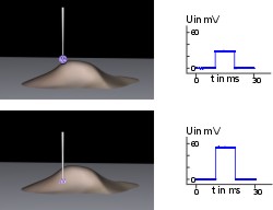
We have developed a novel type of an SICM which deviates from the original
method of Hansma et al. (1) in several respects: The stability of the system
against DC shifts was improved by controlling the approximation of the
electrode to the membrane surface by using short current pulses to the
monitor resistance changes which indicate a close approximation of the
electrode tip to the insulating membrane. Furthermore, we introduced a
"backstep"-mode, which retracts the electrode before lateral movements,
thus preventing the electrode tip from running into possibly overhanging
parts of membrane, especially at the interfaces between culture dishes and
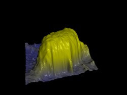
cells (5). The pulse-mode SICM can now be applied to study movements and
volume changes of complete nerve and glial cells in the range of 20 minutes
to hours with lateral step sizes of 0.5 µm and vertical step sizes of 0.1
nm (6).
|